

CeNS Colloquium
Webinar
Date: 09.07.2021, Time: 15:30h
Multidimensional electron microscopy: An interdisciplinary platform for materials and soft matter characterization down to subatomic scales
Prof. Knut Müller-Caspary, Forschungszentrum Jülich and RWTH Aachen
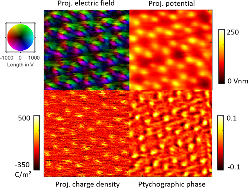
With a spatial resolution below 50 pm, Scanning Transmission Electron Microscopy (STEM) has crossed the border to open the view inside atoms. In fact, STEM is nowadays capable of mapping subatomic electric fields and charge densities which augments its established strengths such as atomic-scale chemical characterisation employing high-angle scattering, X-ray and electron spectroscopy. Recently, ultrafast electron detection paved the way to transform electron microscopy into a multidimensional nanolaboratory that allows for the combination of, e.g., simultaneously acquired real and momentum space measurements at a single-event detection basis.
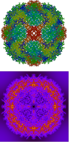
This presentation will briefly introduce established concepts of quantitative STEM to measure strain and composition down to the atomic scale using semiconductor and ferroelectric nanostructures as prominent examples. It will then highlight the concept of momentum-resolved STEM in which a full diffraction pattern is recorded at each scan position using cameras running at frame rates of several kHz, yielding a 4D data set. Firstly, focus is on first moment imaging to map atomic electric fields in 2D materials such as WSe2 in Fig. 1, which also contains the electrostatic potential and charge density derived from the electric field. We then turn to more recent applications of first moment imaging, such as mapping polarisation-induced electric fields in nitride semiconductors. Secondly, the talk deals with electron ptychography based phase retrieval and compares the results with first moment data from the same acquisition. We pick up the excellent dose efficiency of ptychography to motivate the study of dose-sensitive specimen as exemplified for a protein in Fig. 2. Thirdly, the presentation reports focus-variation 4D STEM which allows the retrieval of the 3D shape of nanostructures. Finally, prospects and pilot experiments for time-resolved imaging employing detectors with ps time resolution will be presented, focusing on applications in magnetic dynamics and spectroscopy.
The presentation closes with a perspective view on interdisciplinary research activities in the field of TEM at the faculty of pharmacy and chemistry at LMU, starting from September 2021.
 |
 |